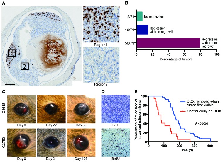
Figure 5. MYCN-independent retinoblastomas emerge in the absence of DOX.
(A) BrdU analysis showing regions with extensive proliferation in Rb/TET-MYCN retinae in the DOX-OFF condition 27 days after DOX removal, while other regions were nonproliferative. Black scale bar: 500 μm; white scale bars: 25 μm. Five of twelve eyes examined showed pockets of proliferation, with one section examined per eye. (B) Histogram showing the proportion of eyes with no tumor regression upon DOX removal, with regression but no regrowth, and with regression followed by DOX-independent tumor regrowth. (C) Photographs showing tumor regression upon DOX removal and then reappearance in the anterior chamber of the eye in 2 animals. (D) BrdU analysis showing a high proliferation rate in DOX-independent returned tumor. Scale bars: 50 μm. (E) Kaplan-Meier curves showing the time to development of late-stage retinoblastoma filling the eye. Rb/TET-MYCN mice were either maintained continuously on DOX (n = 17), or had DOX removed from their diet when the retinoblastoma was first externally visible in the eye (n = 42), usually with tumor evident in the anterior chamber. The P value shown was determined by log-rank test.

