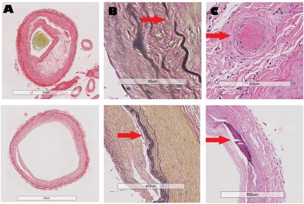
Figure 1. Examples of the arterial characteristics evaluated in this study.
Legend: Column A, Red Sirius staining. Top: Evidence of necrotic core separated from the lumen by a thin fibrous cap in the context of eccentric intima thickening. Bottom: Concentric intima hyperplasia without evidence of cholesterol deposition. Column B, Van Gieson staining. Top: Evidence of gap in the internal elastic lamina (arrow). Bottom: Duplication of the internal elastic lamina (arrow). Column C, H&E staining. Top: evidence of vasa vasorum (arrow) in the adventitia of an artery. Bottom: Evidence of confluent arterial calcification in absence of cholesterol deposition or atheroma.

