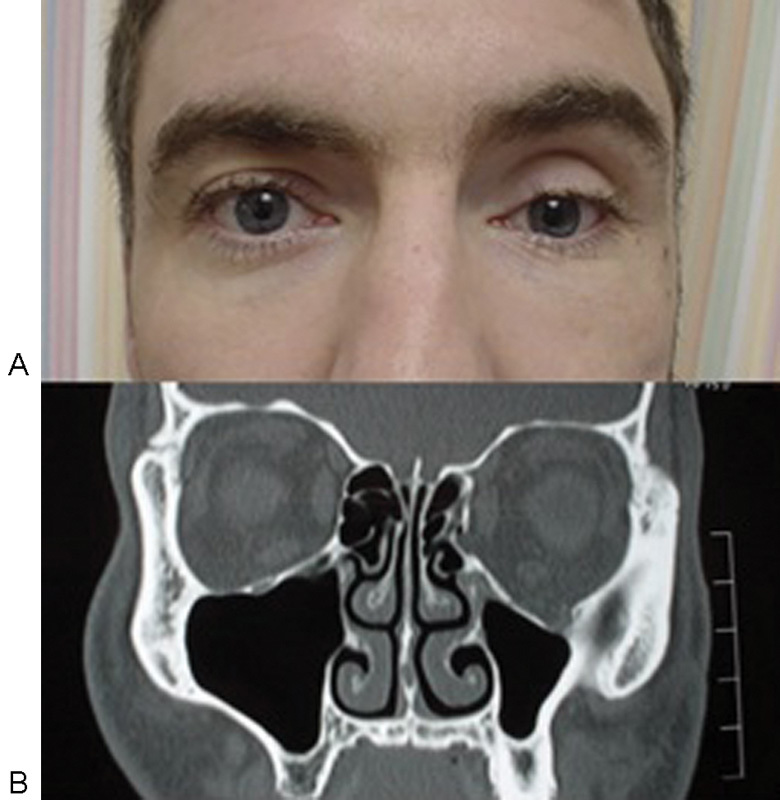
Fig. 4.

Photograph (A) showing evidence of enophthalmos on the left that is suggested clinically by the smaller palpebral fissure and deepened superior sulcus. Coronal computed tomography image (B) showing an inferior orbital wall fracture and an increase in the orbital volume compared with the noninjured right orbit.
