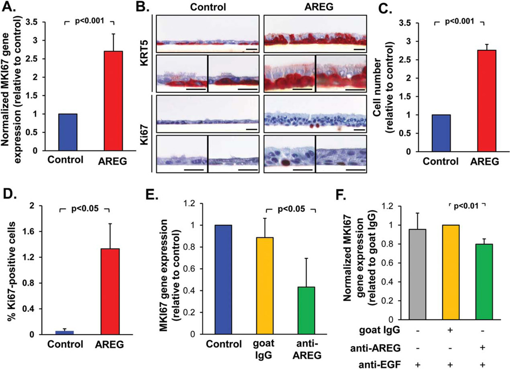
Figure 4.
AREG promotes airway basal cells (BC) proliferation and hyperplasia. (A): Expression of the MKI67 gene in the epithelium derived from airway BC after 28 days of air-liquid interface (ALI) in the presence of AREG relative to control group (mean±SD). (B): Morphology and immunohistochemistry of samples derived as described in A for BC marker KRT5 and proliferation marker Ki-67. Shown are three separate areas of the ALI cultures for each of marker; scale bars-20 µm. (C): Total cell number (normalized to the control group) and (D) % Ki-67+ cells in the samples derived from BC as described A in three randomly selected fields (mean ± SD). (E). MKI67 gene expression in the epithelium derived from BC during 14 days in ALI ± neutralizing anti-AREG antibody or goat IgG isotype control (both 1 µg/ml; normalized to control group; mean ± SD). (F): MKI67 gene expression in the airway epithelium derived from BC treated with EGF-conditioned media (EGF-CM; see text) ± neutralizing anti-AREG antibodies (1 µg/ml), and neutralizing anti-EGF antibody (0.5 µg/ml); normalized to the control IgG group (mean ± SD); for more data see Supporting Information Figure S4. All panels represent data derived from ≥3 independent experiments. Abbreviations: AREG, amphiregulin; EGF, epidermal growth factor.

