
Figure 1. In vivo isolation of the sciatic nerve and fast‐twitch medial gastrocnemius (MG), extensor digitorum longus (EDL) and tibialis anterior (TA) muscles and the slow‐twitch soleus (SOL) muscle for isometric contractile force recording.
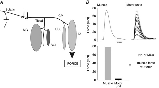
A, all nerves except the sciatic and its tibial and CP nerve branches to the MG and SOL muscles and to the EDL and SOL muscles were denervated. Each of the muscles was attached to a force transducer for the recording of maximal muscle twitch contractile force and, in response to all‐or‐none increments in stimulus intensity on the sciatic nerve, the progressive recruitment of single motor units (B). The number of motor units (MUs) was obtained from the ratio of muscle and MU forces as described in the text.