
Figure 1. Histological verification of the electrode placement.
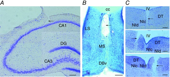
A, lesion of the LFP recording electrode aimed at the hippocampal CA1 (composite image). B, injection site lesion in the MS (composite image). C, top, trace of the muscimol injection in the NI. Middle, electrical stimulation electrode misplacement in a neighbouring area to the NI. Bottom, electrical stimulation electrode lesion inside the boundaries of the NI. [Colour figure can be viewed at wileyonlinelibrary.com]