
Figure 5.
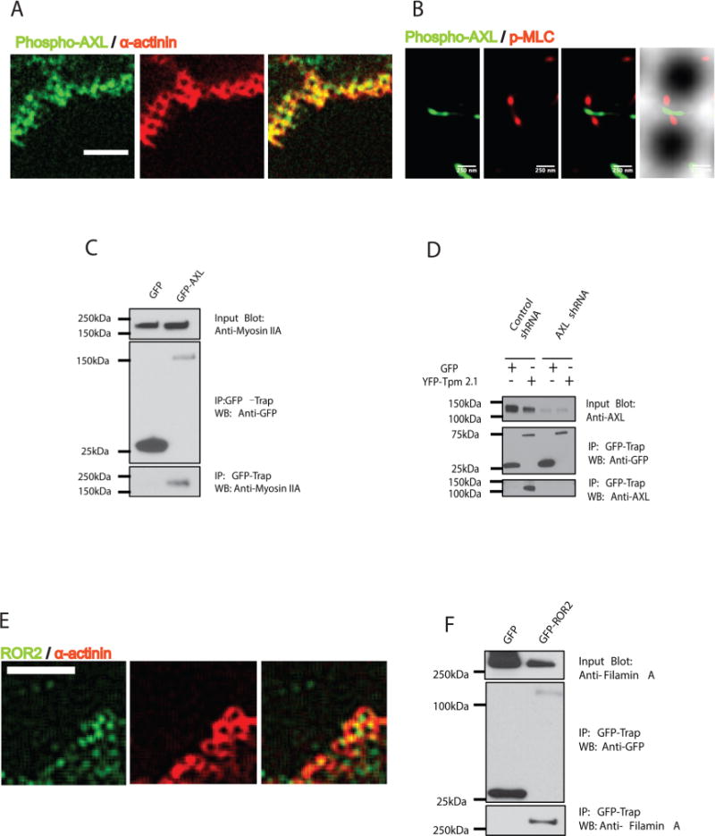
AXL and ROR2 associate with components of contraction units. (A) Phospho-AXL (green) colocalized around pillars together with α-actinin (red) at the cell periphery during initial spreading (15 min; scale bar 5 μm). (B) Phospho-AXL (green) overlaps p-MLC (red) in super-resolution images (3B analysis) in contractile regions at cell edges. (C) GFP-AXL transfected HFF cell lysates were immunoprecipitated with GFP nanotrap beads, followed by immunoblotting with anti-Myosin IIA antibody. (D) YFP-Tpm 2.1 transfected normal HFF cell or AXL knockdown cell lysates were immunoprecipitated with GFP nanotrap beads, followed by immunoblotting with anti-AXL antibody. (E) ROR2 (green) overlaps with α-actinin around the pillars at the cell periphery during initial spreading time (15 min; scale bar 5 μm). (F) GFP-ROR2 transfected HFF cell lysate was immunoprecipitated with GFP nanotrap beads, followed by immunoblotting with antifilamin A antibody.