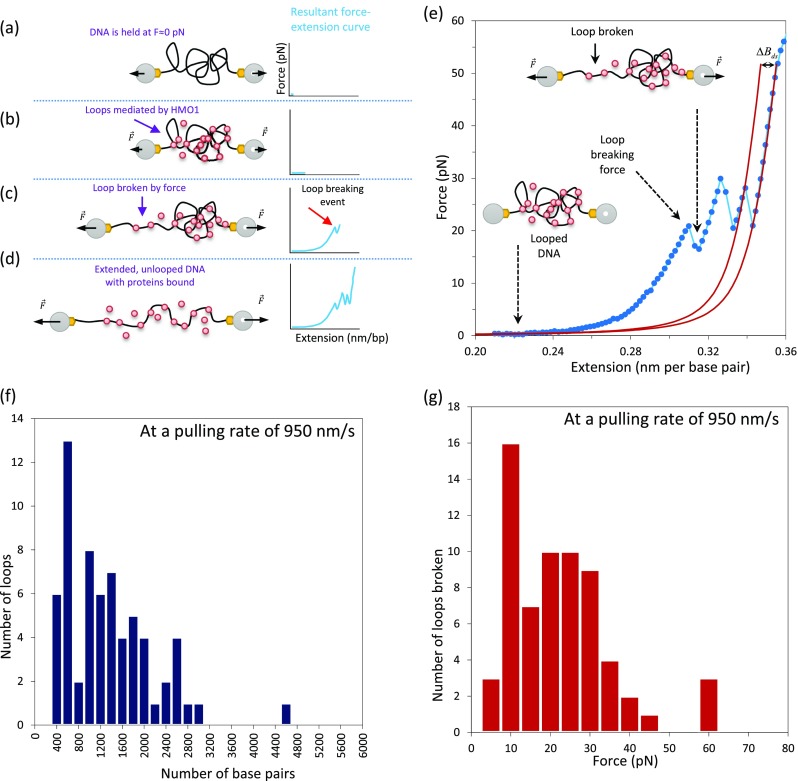
Fig. 10.

Protein–DNA loop formation as a mechanism for DNA compaction. a–d Schematic illustrating the formation and breaking of loops. When the DNA is held at low forces, HMO1 proteins are able to mediate and stabilize loops and the force–extension curve is relatively flat (b). Here, the blue line represents the force–extension curve. In contrast, when the DNA is extended further, the force–extension curve shows jumping events, revealing the breaking of loops mediated by HMO1 (c). As the DNA is further extended, unlooped DNA with proteins bound is stretched (d). e Force–extension curves for phage λ DNA in the presence of 0.3 nM HMO1. Each jump illustrates a loop-breaking event. Fitting is to the WLC model (solid red lines). Loop size is estimated by measuring the contour length change over the force jump. f Loop sizes. The most probable loop size is between 400 and 600 bp. g Loop breaking forces. The most probable loop breaking force is between 10 and 15 pN at this pulling rate of 950 nm/s. (Adapted from Murugesapillai et al. 2014)
