Fig. 3.

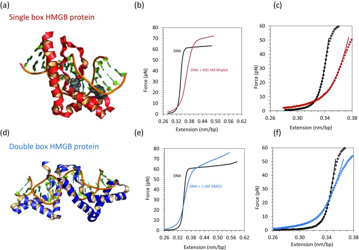
Binding of Nhp6A and HMO1 proteins to λ DNA characterized by optical tweezers. a Solution structure of the yeast single box Nhp6A protein bound to DNA with intercalating amino acid side chains shown as gray space-filled atoms (PDB code: 1J5N). b Force–extension curves are shown for phage λ DNA in the absence (black) and presence (red) of the single box Nhp6A protein. c Fits to the WLC model in the absence (black) and presence (red) of Nhp6A. d Solution structure of a double box HMGB protein bound to DNA (PDB code: 2GZK). e Force–extension curves are shown for phage λ DNA in the absence (black) and presence (blue) of the double box HMO1 protein. f Fits to the WLC model in the absence (black) and presence (blue) of HMO1. (Adapted from McCauley et al. 2013; Murugesapillai et al. 2014)
