
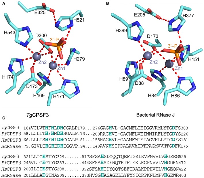
Figure EV4. Conservation of catalytic residues in Toxoplasma gondii CPSF3.
-
A, BCoordination of Zn atoms at the catalytic sites of the T. gondii CPSF3 model (A), same model as in Fig 3A, and Streptomyces coelicolor RNase J (B), PDB: 5A0T. The position of the phosphate of the 3′‐mRNA at the cleavage position is shown for reference. Key interactions are shown as red dashed lines.
-
CSequence alignment of regions constituting the catalytic sites of CPSF3 homologous proteins of Toxoplasma gondii (Tg), Plasmodium falciparum (Pf), Homo sapiens (Hs) and of RNase J from Streptomyces coelicolor (Sc).