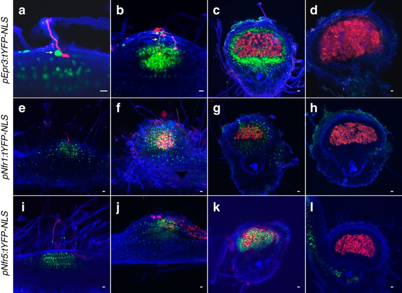
Figure 5. Temporal and spatial expression of Epr3 and Nfr promoter reporter constructs.
Images of the infection process from the root hair infection stage (a,e,i) to nodule maturity (d,h,l) were collected from sections/whole mounts of transgenic roots inoculated with M. loti MAFF303099 DsRed. (a–d) pEpr3:tYFP-NLS, (e–h) pNfr1:tYFP-NLS and (i–l) pNfr5:tYFP-NLS. White and yellow arrows show specific Epr3 expression in epidermal and outer cortical cells adjacent to infection threads. Scale bars, 20 μm.

