
Figure EV1. PERK activity in vivo in the mouse model.
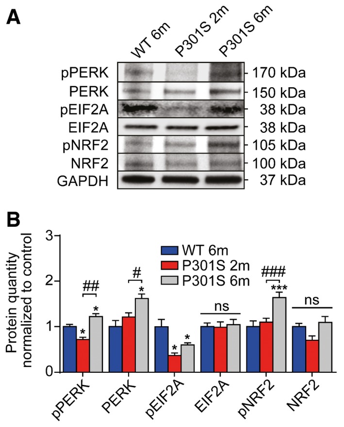
- Western blots of whole‐brain extracts of mice (wild‐type mouse: WT; P301S transgenic mouse: P301S, 2 months of age: 2m, 6 months of age: 6m; n = 3 per group) were done with antibodies against phosphorylated PERK (pPERK), total PERK, phosphorylated EIF2A (pEIF2A), total EIF2A, phosphorylated NRF2 (pNRF2), and total NRF2. GAPDH was used as loading control.
- Quantification of (A). Data are mean + SEM. Statistical analysis was one‐way ANOVA followed by Student–Newman–Keuls post hoc test, *P < 0.05, ***P < 0.001 versus WT 6m; # P < 0.05, ## P < 0.01, ### P < 0.001 versus P301S 2m, ns: not significant.