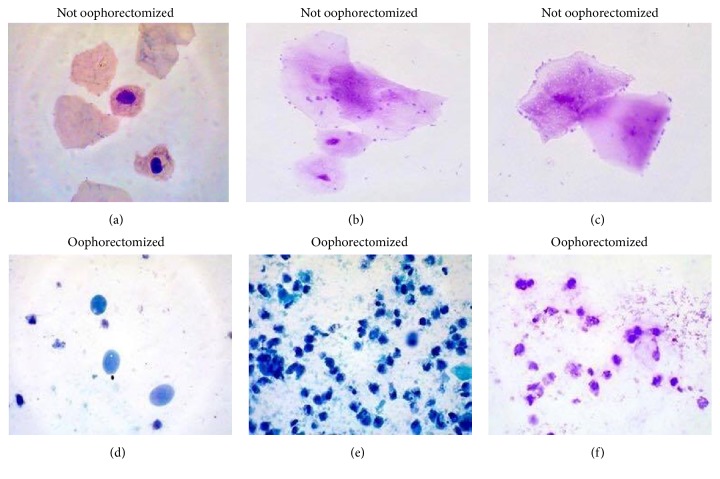
Figure 3.
Histology in rats with and without oophorectomy. Vaginal smear from not oophorectomized animal. (a) Anuclear squamae are seen. Papanicolaou stain ×40. (b) and (c) Diff-Quik stain. (d) Parabasal epithelial cells are seen in vaginal smear from an oophorectomized rat, Papanicolaou stain ×40. (e) and (f) Diff-Quik stain.

