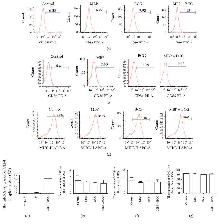
Figure 4.
Effect of the combination of MBP and BCG on the phenotypic maturation of DCs from TLR4−/− mice. Splenic DCs from TLR4−/− mice (1 × 10⁶ cells/mL) were cultured with MBP, BCG, or the combination of MBP and BCG for 48 h and were analyzed using flow cytometry. (a–c) DCs collected from the different groups were stained with the following antibodies: FITC-conjugated anti-CD80, PE-conjugated anti-CD86, and APC-conjugated anti-MHC class II. The percentage of positive cells is shown in a flow cytometry histogram. (e–g) The percentage of CD11c+ cells expressing each surface molecule is expressed as the mean ± standard deviation and is shown in a bar graph. Data are derived from three independent experiments. ∗P < 0.05 is considered to indicate statistically significant differences, compared with control group derived from WT mice. (d) The mRNA level of TLR4 in spleen tissue from TLR4−/− mice or from WT mice was measured with RT-PCR.

