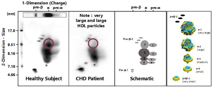
Figure 1.
Two dimensional gel electrophoresis patterns of HDL particles from a normal subject (far left) and a patient with premature coronary heart disease (CHD) (second from left) along with two depictions of the position (middle right) and the potential structure (far right) of apoA-I containing HDL particles are shown. On the gel patterns the particle size in nm (diameter) is plotted on the vertical axis and the electrophoretic mobility (preβ, α, and preα) is plotted on the horizontal axis. The CHD patient clearly has a marked reduction in the apoA-I concentration in very large α-1 HDL.

