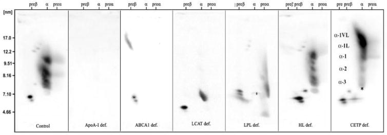
Figure 2.
Two dimensional gel electrophoretic patterns of apoA-I containing HDL particles (from left to right) are shown from a control subject, an apoA-I deficient patient (no particles), a TD patient (only preβ-1 HDL), an FLD patient (preβ-1 and α-4 HDL, with some larger discoidal fusion particles), a LPL deficient patient with lack of large α HDL, an HL deficient patient with decrease in α-2 HDL, and a patient with CETP deficiency with an excess of abnormal very large HDL particles.

