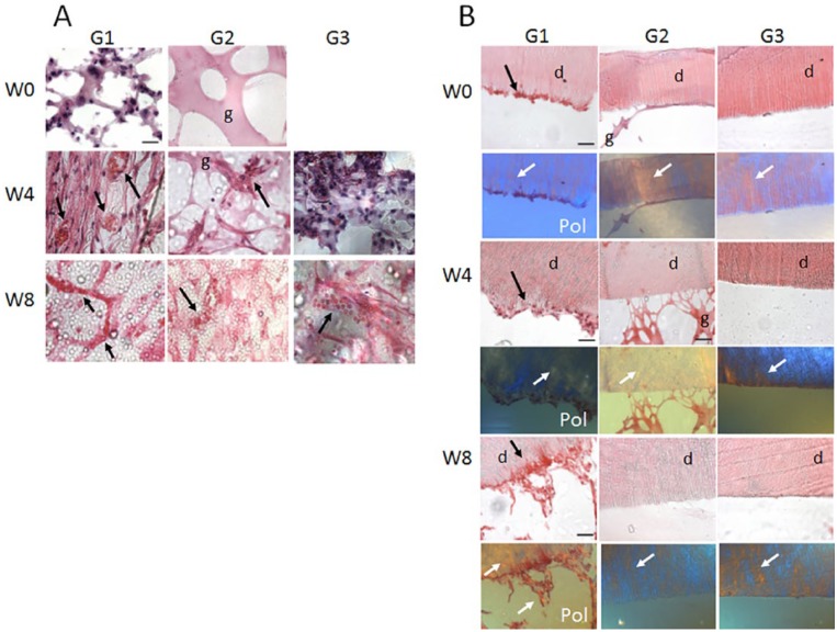
Figure 5.
Bioengineered dental pulp vascularization and interactions with inner dentin surface. (A) High-magnification images of the pulp center of representative in vivo implanted G1, G2, and G3 constructs. G1, human dental pulp stem cell (hDPSC)/human umbilical vein endothelial cell (HUVEC)–encapsulated gelatin methacrylate (GelMA) constructs exhibited high cellularity at week 0, as well as increased cellularity and host red blood cell (RBC) circulation at 4 and 8 wk. G2 acellular GelMA samples exhibited a degree of host cell infiltration at 4 wk and disorganized vasculature formation at 8 wk of implantation. G3, empty tooth root segment (RS) exhibited some host cell infiltration at 2 wk and less well-organized host blood vessel formation at 8 wk compared with G2 constructs. (B) The inner dentin surface of G1, hDPSC/HUVEC-encapsulated GelMA constructs showed hDPSC/HUVEC cell attachment after 1 wk of in vitro culture (week 0) and increased cell attachment over in vivo implantation time (week 4, week 8). Polarized light imaging revealed increased collagen deposition and organization at the inner dentin surface over in vivo implantation time and increased formation and length of cellular extensions into the dentin tubules, indicative of reparative dentin formation and tooth revitalization. G2, acellular tooth root constructs showed GelMA attachment at week 0 and week 4 but no remaining GelMA present at week 8. Although host cells infiltrated into acellular GelMA construct pulp centers, no host cell attachment to inner dentin surface was observed in these constructs. Similarly, G3, empty tooth RSs did not show cell attachment to inner dentin surface at any of the times investigated.

