
Figure 2.
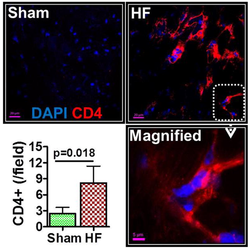
Representative confocal images of CD4 immunostained hearts from sham-operated and HF mice (8 w post-MI). CD4 positivity is indicated by red fluorescence; DAPI-labeled nuclei are blue. A magnified image is shown in the bottom right, and group quantitation of cardiac CD4+ cell abundance in the lower left.