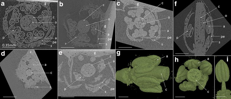
Fig. 2.

X-ray µCT XY cross section image of Arabidopsis single buds at different developmental stages. Flower staging based on [30]; Flower stages 11 (a, f), 10 (b), 9 (c, d), 13 (e) showing individual organs, pollen and ovules. 3D representative images (g, h) of bud, and of an isolated anther at flower stage 11 (i). Within the images; carpel (c), anther (a), petal (pe), sepal (s), ovule (o), pollen (p), glue to attach bud to glass needle (g). Scale bar is 0.35 mm
