
Fig. 2.
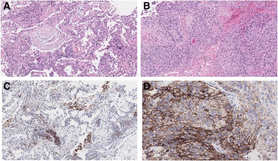
Histological assessment of different lung pathologies. Hematoxylin and eosin staining revealed organizing pneumonia with fibroblast plugs and inflammatory cells in the RML (a), and moderately differentiated squamous cell carcinoma in the RLL (b). Magnification, ×20. IHC staining for PD‐L1 expression shows the presence of PD-L1 positive intra-alveolar macrophages in the RML (c) and strongly positive PD-L1 expression on the surface of 90% of viable tumor cells (TPS ≥50%) (d). Magnification, ×100