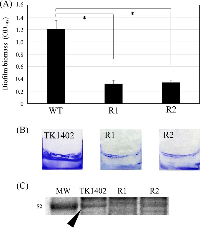
FIG 1.
Biofilm formation by wild-type TK1402 (WT) and two spontaneous TK1402 CAM-resistant strains, TK1402CAMR1 (R1) and TK1402CAMR2 (R2). (A) H. pylori biofilms formed on glass coverslips after 3 days were quantified by crystal violet (CV) staining and ethanol elution. The result is expressed as the means ± 1 standard deviation from at least three independent experiments. (B) CV-stained biofilm of H. pylori strains grown on the surfaces of glass coverslips in brucella-FCS broth. (C) Protein profiles of OMV from wild-type TK1402 (TK1402), TK1402CAMR1 (R1), and TK1402CAMR2 (R2). The approximate position of the 52-kDa band in the wild type is shown by an arrowhead. An asterisk indicates significant difference (P < 0.05) relative to the level of wild-type biofilm biomass. MW, molecular weight in thousands.

