FIG 4.

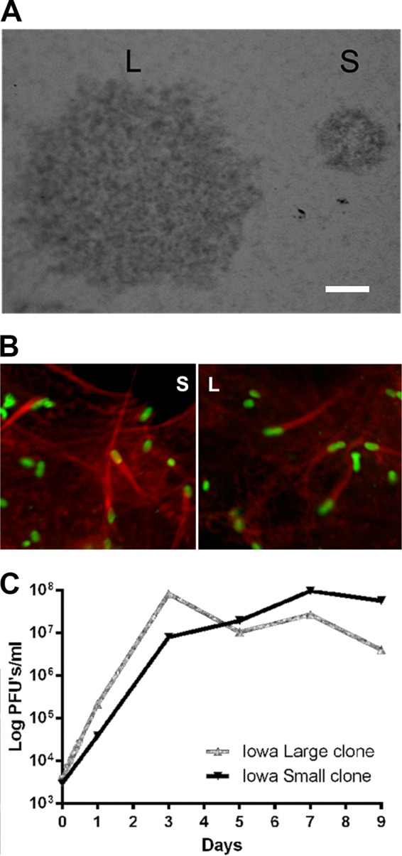
R. rickettsii Iowa low-egg-passage large- and small-plaque morphologies. (A) Vero cell monolayers infected with a low-passage-number (4 EP) Iowa strain revealed two distinct plaque types. A large-plaque variant (L) and a small-plaque variant (S) were subsequently cloned and expanded for further study. (B) Actin polymerization by the L and S variants. Monolayers of Vero cells were infected with the Iowa strain L and S variants for 24 h. Rickettsiae were detected by indirect immunofluorescence using monoclonal antibody 13-2 (1) to rOmpB; F actin was stained with 10 U/ml of rhodamine phalloidin. Bar, 1 mm. (C) Growth curve of the Iowa strain L and S variants. Rickettsiae were grown in Vero cells at 34°C, and samples were taken for cell disruption and for replating the lysates to enumerate the PFU. Data points are the means from three replicates. Error bars representing the standard errors of the mean (SEM) are not visible under the symbols.
