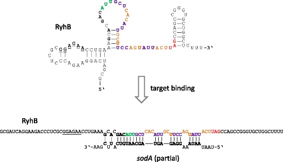
Fig. 6.

Overview of the secondary structures formed by RyhB for the molecule on its own (top) and after binding to a target RNA, like sodA (bottom). Structures are taken from [99]. Individual bases have been highlighted. Underlined, putative Shine-Dalgarno sequence; green, start codon; violet/orange, individual codons along the frame; red, stop codon, bold; bases involved in hybridization to the sodA-target
