
FIG 8.
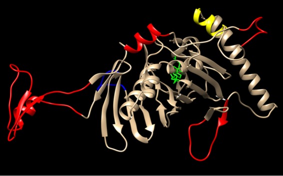
Potential RIDα binding site includes several unique ORP1L insertions. A three-dimensional model of human ORP1L-ORD (NCBI accession no. NP_542164.2) was generated using the structure of the yeast ORP homologue Osh4 as a template (PDB code 1ZHY) (72). Overlay was performed using UCSF Chimera Molecular Modeling software (http://www.cgl.ucsf.edu/chimera/). The ORD backbone is indicated in gold, the α7 helix in yellow, and the bound cholesterol ligand in green. The RIDα binding site has been mapped to ORP1L-ORD residues 755 to 923 encompassing the highly conserved α7 helix and four of the unique ORP1L insertions highlighted in red (15). A fifth unique insertion outside the RIDα binding site is highlighted in blue.