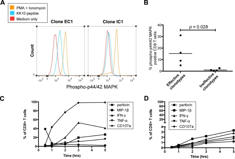
FIG 4.
TCR signal transduction and cytokine kinetics. (A) A representative flow cytometry histogram of phosphorylated protein forms of p44/42 MAPK examined by phosphoflow cytometry at the 10-min time point following stimulation with KK10 peptides or PMA and ionomycin of EC1 and IC1 CD8+ T-cell clones. (B) Comparison of phosphorylation of p44/42 MAPK between effective and ineffective CD8+ T-cell clones was made with the Mann-Whitney test. (C and D) Functional cytokine and chemokine expression was examined by flow cytometry after incubation with KK10 peptide-loaded HLA-B*2705-expressing GXR cells for 0.5 h, 1 h, 2 h, 3 h, and 5 h for effective CD8+ T-cell clone EC1 (C) and ineffective CD8+ T-cell clone IC1 (D).

