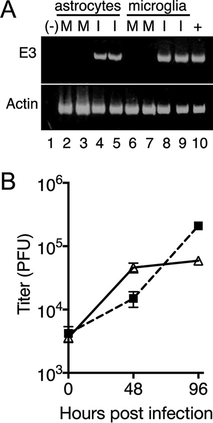
FIG 4.

MAV-1 infects cultures enriched for astrocytes and microglia. Primary astrocytes and microglia were infected with MAV-1 at an MOI of 5 or mock infected and harvested at 48 hpi (A) or at the times shown (B). (A) cDNA was prepared and assayed by PCR for E3 and actin, as indicated. −, negative control (water); +, positive control (cDNA from mRNA from MAV-1-infected 3T6 cells 48 hpi). Lanes 2 to 5, astrocytes; lanes 6 to 9, microglia. M, mock infected; I, infected. Each condition was assayed on duplicate wells of cells from the same primary cell preparation. The experiment shown is representative of six experiments from independent primary cell preparations. (B) Cells and supernatants were harvested and titrated on mouse 3T6 cells. Each point indicates the mean and standard deviation of three plaque assay plates. The experiment shown is representative of four experiments from two independent primary cell preparations. Triangles, astrocytes; squares, microglia.
