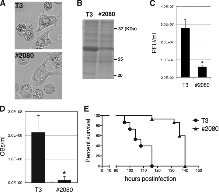
FIG 1.
Characterization of a novel BmNPV FP mutant. (A) Light microscopic observations at 3 dpi of BmN-4 cells infected with T3 or #2080. (B) POLH expression. The cell lysate of BmNPV-infected cells was subjected to SDS-PAGE at 3 dpi, and a gel stained with Coomassie brilliant blue (CBB) is shown. (C) Cellular BV production. The BV titer at 3 dpi was determined by plaque assay. Data are presented as the mean ± standard deviation (SD) from triplicate experiments. *, P < 0.05, Student's t test. (D) Larval OB production. The numbers of OBs released at 4 dpi in the hemolymph of BmNPV-infected B. mori larvae were counted (n = 5). Data are presented as the mean ± SD. *, P < 0.05, Student's t test. (E) Survival curves for B. mori larvae infected with T3 or #2080. The LT50s of T3 and #2080 were 108 and 140 h, respectively (n = 15).

