
FIG 3.
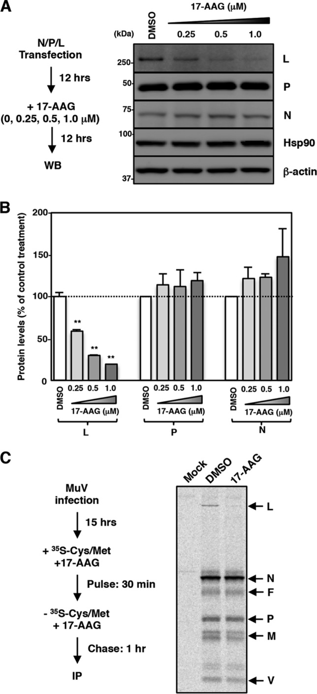
MuV L protein is a client of Hsp90. (A and B) 293T cells were cotransfected with three plasmids, pCAGGS-N, -P, and -L, and the medium was replaced with fresh medium containing the indicated concentrations of 17-AAG at 12 h posttransfection. At 24 h posttransfection, the cell lysates were subjected to immunoblotting with the indicated antibodies (A). The relative band intensities of each viral protein were normalized by the β-actin level, and the relative expression levels of each viral protein based on the levels of DMSO-treated cells are shown in panel B. (C) Vero cells were infected with MuV at an MOI of 5.0. At 15 hpi, the cells were radiolabeled for 30 min with [35S]methionine-cysteine and incubated in nonradioactive medium for 1 h before lysis and analysis by immunoprecipitation with an anti-MuV antibody. 17-AAG was incubated at 0.5 μM during the pulse and chase periods. WB, Western blotting; IP, immunoprecipitation.