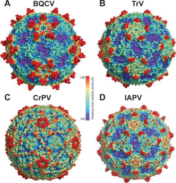
FIG 1.

Comparison of virion structures of BQCV, TrV, CrPV, and IAPV. Molecular surfaces of BQCV (A), TrV (B), CrPV (C), and IAPV (D) virions are rainbow-colored based on their distance from the virion center. Depressions are shown in blue and protrusions in red.
