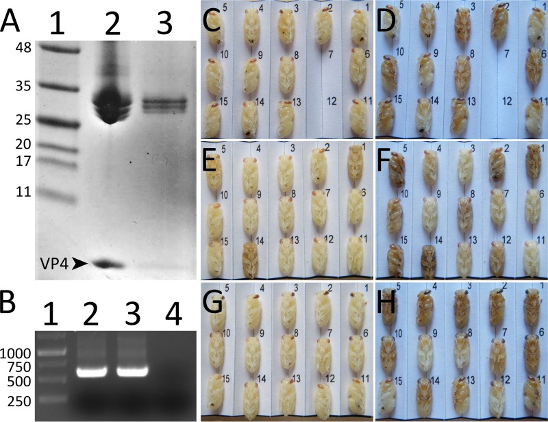
FIG 7.
BQCV crystals contain VP4 subunits and the crystallized virus is infectious. (A) Polyacrylamide gel electrophoresis of capsid proteins of BQCV. Lane 1, marker; lane 2, purified BQCV; lane 3, BQCV dissolved from crystals. Arrowhead and VP4 label indicate the position of capsid protein VP4 (8.1 kDa). Capsid proteins VP1, VP2, and VP3 of BQCV have molecular masses in the 25- to 35-kDa range. (B) Agarose gel electrophoresis of PCR fragments obtained from reverse-transcribed RNA isolated from pupae injected with native BQCV (lane 2), BQCV dissolved from crystals (lane 3), and mock-infected with PBS (lane 4). Please see Materials and Methods for details. Lane 1, DNA ladder. (C to H) Images of pupae injected with BQCV dissolved from crystals (C and D) or native virus (E and F) or mock infected with PBS (G and H). The pupae were imaged 1 day (C, E, and G) and 5 days (D, F, and H) after the injection. The pupae injected with virus (C to F) developed slower than the mock-injected pupae (G and H), as shown by the delay in color development of the eyes and the darkening of the body 5 days postinfection. Two pupae missing in the panels (C and D) were accidentally destroyed during imaging.

