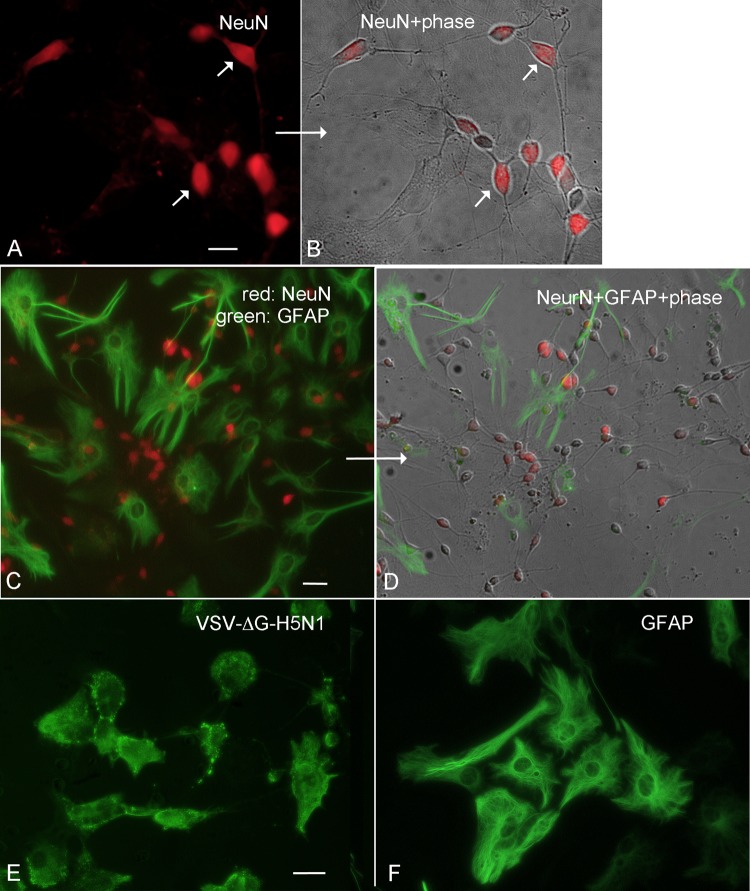
FIG 2.
Identification of neurons and astrocytes. (A) Neuronal nuclei are immunostained red for the neuronal antigen NeuN (short arrows). (B) Neurons with 2 to 4 thin dendritic or axonal processes contain NeuN-positive nuclei. (A and B) Bar, 20 μm. (C) Neurons are labeled by NeuN immunostaining (red), and astrocytes are labeled green by GFAP immunostaining. (D) In the same field shown in panel C, phase contrast shows that phase-bright neurons and astrocyte immunostaining overlap. (C and D) Bar, 40 μm. (E) Astrocytes 2 days after inoculation with VSVΔG-H5N1. Astrocyte processes have withdrawn, and GFAP immunoreactivity is no longer fiber-like, as seen in normal astrocytes (C and F), but has become more globular, indicating cell cytolysis. (F) Normal astrocytes immunolabeled with GFAP show long, thin GFAP staining. (E and F) Bar, 40 μm.

