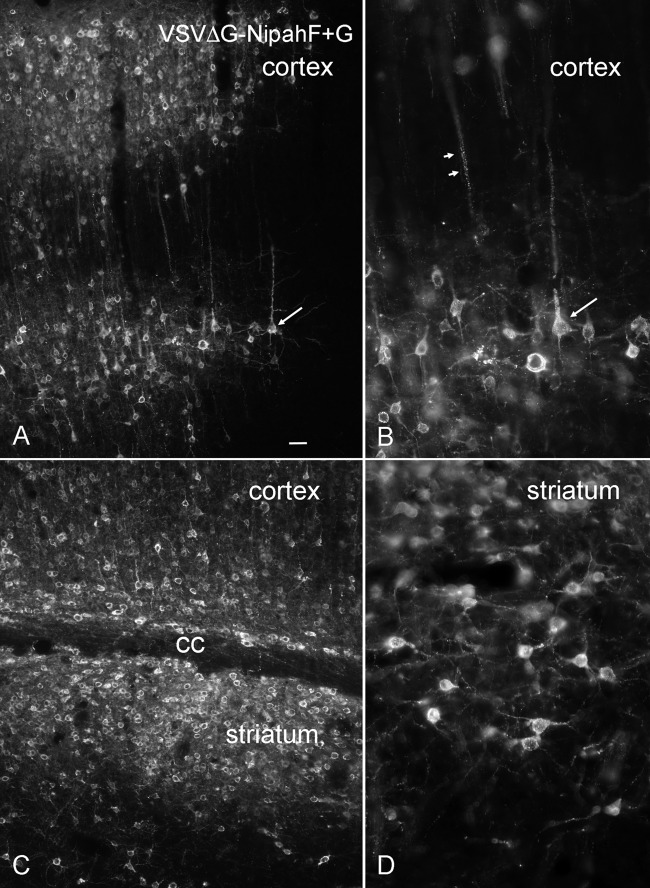
FIG 9.
VSVΔG-Nipah F+G in the brain. (A) At 1 day after a small, 0.4-μl intracortical injection, VSVΔG-Nipah F+G had spread to a large number of neurons. The arrow indicates a neuron with pyramidal cell morphology. Bar, 22 μm. (B) Higher magnification of panel A. Arrows point to the same neuron in panels A and B. (C) Virus spread across the corpus callosum (cc) into the striatum. (D) Higher magnification of infected striatal cells. In both the striatum, as shown, and the cortex (B), most infected cells had the morphology of neurons. No infection was seen in the contralateral cortex.

