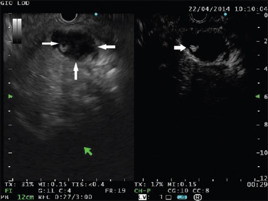
Figure 1.

A cystic lesion is seen in the pancreatic head containing solid components with mixed echogenicity (left panel, thin arrows). At contrast harmonic-endoscopic ultrasound, a small area of hyperenhancement is clearly visible (right panel, arrow). This finding is highly suggestive of malignancy
