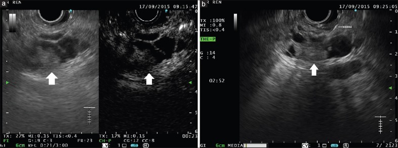
Figure 3.
(a) A complex multiloculated cystic lesion is visible in the pancreatic head, containing a hypoechoic solid component (left panel, arrow). At contrast harmonic-endoscopic ultrasound, the solid component appears hyperenhanced (right panel, arrow), as well as septae and cystic wall. (b) As the contrast harmonic-endoscopic ultrasound finding is highly suggestive of malignancy, selective endoscopic ultrasound-fine-needle aspiration is performed

