Figure 3.
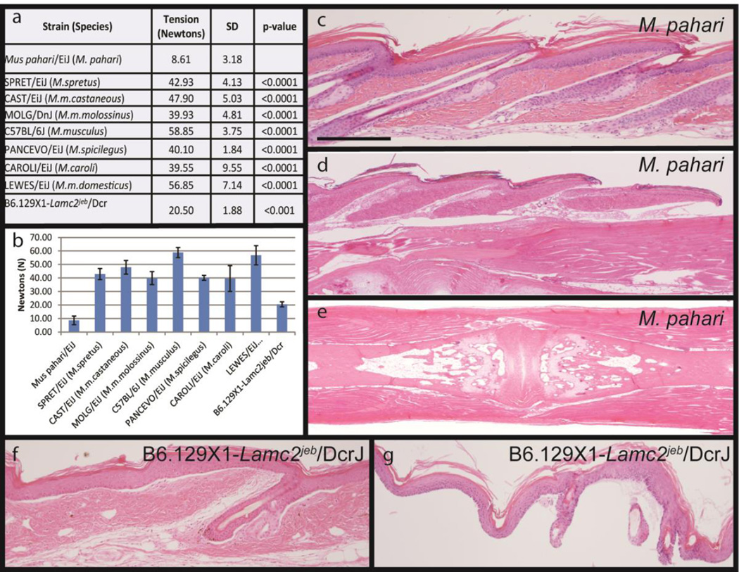
The tail tension assay removes the tail skin and provides a quantifiable phenotype for comparison between Mus species and different mutant mouse strains that have somewhat similar fragile skin (a,b). Tail tension testing was used to measure the force, in Newtons, required to strip the skin from the mouse tail. Tension testing failed to remove skin from SPRET/EiJ, CAST/EiJ, MOLG/DnJ, PANCEVO/EiJ, CAROLI/EiJ, LEWES/EiJ or C57BL/6J within the limits of the equipment. Mus pahari skin consistently separated at a low force, typically 7–12 Newtons, with no apparent age or sex differential. Histology is required to determine where in the skin the separation occurs. For Mus pahari the separation occurs below the hypodermal fat layer. Skin removed from the tail (c) has an intact epidermis and dermis but the separation occurred below the fat layer (arrows). At the point where the full thickness skin separates as it is pulled off (d, arrow) a space forms between the dermis and underlying ligaments surrounding the coccygeal vertebrae. The remaining coccygeal vertebrae have only muscle and ligaments remaining (e). By contrast, mice with junctional epidermolysis bullosa (B6.129-Lamc2jeb/jeb) have separation at the level of the basement membrane (f). In the tail tension assay there is complete dermal-epidermal separation (g). H&E stain. p>value was calculated by Student’s t-test. Scale bar=200uM

