
Figure 4.
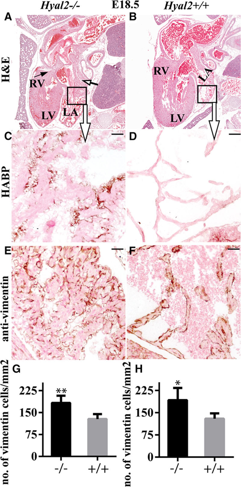
Histological analysis of Hyal2−/− and control hearts at embryonic day (E) 18.5. Paraffin sections from E18.5 embryos were stained or used for immunohistochemistry. A and B, Images of hematoxylin and eosin–stained embryo sections revealed an enlarged atrium (open arrow) and the presence of fibrosis (arrow) in the Hyal2−/− heart (A) compared with the control (B) heart. (Hyal2−/−, n=8; control, n=6). C–F, Enlarged view of the area in the box in (A) and (B) stained for hyaluronan (HA). C and D, Images showing increased HA (brown) in the Hyal2−/− atrium (C) compared with the control atrium (D; n=3). E and F, Detection of vimentin-positive cells (brown) revealed an excess in the Hyal2−/− atrium (E) compared with the control (F; n=3). G–H, Semi-quantitative analysis of vimentin-positive cells in the atrium (G; n=3) and ventricle (H; n=4) of Hyal2−/− and control hearts. There were significantly increased numbers of mesenchymal cells in the Hyal2−/− hearts compared with controls. Bar=50 μm. LA, left atrium; LV, left ventricle; and RV; right ventricle. *P<0.05; **P<0.001.