
FIGURE 9.
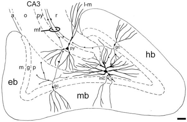
Concluding diagram. Semidiagrammatic drawing of the dentate gyrus and adjacent part of CA3 with focus on the shape of the dendritic arbors of granule cells (gc), mossy cells (mc) and CA3 pyramidal cells (py) and their relation to subdivision boundaries. Axons are not included except for the granule cell axons (mossy fibers, mf), which feature giant terminals synapsing with the thorny excrescences of both mossy and pyramidal cells. The somata and excrescence covered proximal dendrites of mossy cells are confined to the polymorph layer. Special, so-called gm-dendrites (arrows), extend into the granule and molecular layers. Distal dendrites of mossy cells may extend into the subjacent CA3, while dendrites of pyramidal cells seem to respect the border. Abbr.: a, alveus; g, granular layer; eb, exposed blade; gc, granule cell; hb, hidden blade; l-m, lacunar-molecular layer; m, molecular layer; mb, middle blade; mc, mossy cell; mf, mossy fibers; o, oriens layer; p, polymorph layer; py, pyramidal layer/cell; r, radiate layer. Calibration bar, 100 μm.