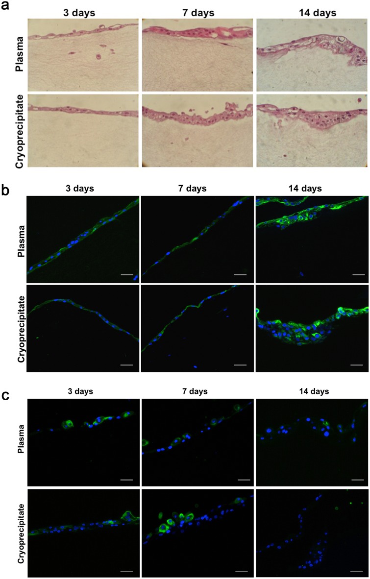
Fig 2. Conjunctival cells grown in and on fibrin scaffold constructs.
(a) Cell seeded constructs derived from plasma or cryoprecipitate were stained with hematoxylin/eosin at days 3 (left), 7 (middle), and 14 (right). The number of epithelial cell layers increased over time. Magnification: X20. (b) Representative microphotographs of CK19 staining in the constructs. Epithelial cells maintained CK19 staining (green) up to day 14 in both plasma (top) or cryoprecipitate (bottom) scaffolds. Cell nuclei are stained with Hoechst dye (blue). Scale bar: 50 μm. (c) Representative microphotographs of HPA staining in the conjunctival constructs. Epithelial cells cultured on the surface of fibrin scaffolds produced mucins (green) at days 3 (left) and 7 (middle), but they did not produce them at day 14 (right). Cell nuclei are stained with Hoechst dye (blue). Scale bar: 50 μm.

