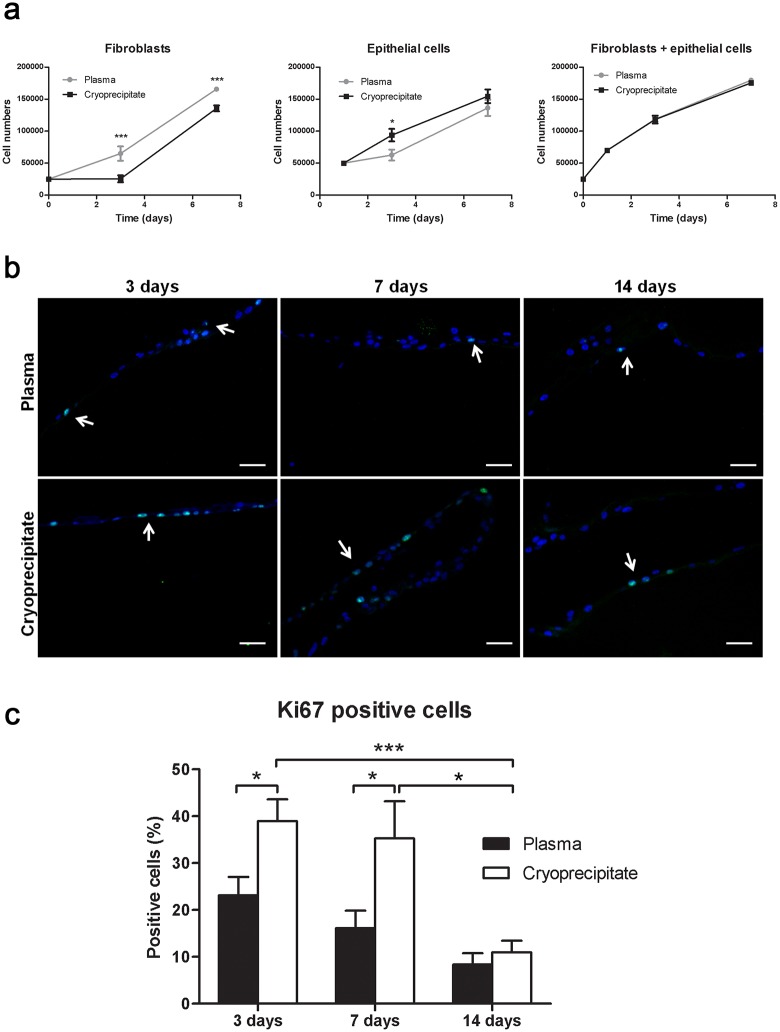
Fig 3. Conjunctival cells proliferated at different rates when seeded in plasma or cryoprecipitate scaffolds.
Proliferation was measured by Ki67 staining (green). (a) Proliferation of fibroblasts alone (left), epithelial cells alone (middle), or both (right) was measured with alamarBlue® assay in four independent experiments. *p ≤ 0.05; ***p ≤ 0.005. (b) Some Ki67-stained cells are indicated by arrows. Nuclei were stained with Hoechst dye (blue). Scale bar: 50 μm. (c) The percent of positive cells for the proliferation marker Ki67 was higher in cryoprecipitate scaffolds than in plasma ones at days 3 and 7. That percent was significantly decreased at 14 days. * p ≤ 0.05; *** p ≤ 0.005.

