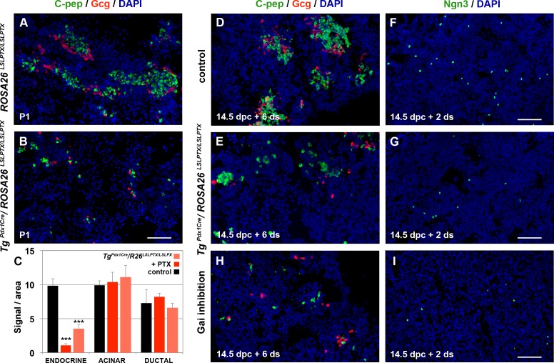
Fig 5. Gαi subunits mediate endocrine pancreas specification.
(A-I) Activation of PTX expression from two alleles of the ROSA26 LSLPTX transgene using the TgPdx1Cre driver resulted in a striking loss of C-pep+ and Gcg+ cells at P1, as shown by immunofluorescence analysis (A, B; Quantitations are provided in S6L Fig). ALI cultures of 14.5 dpc embryonic pancreata expressing PTX from two alleles in Pdx1+ progenitors also resulted in reduced numbers of C-pep+ and Gcg+ cells after 6 d in culture (C, D, E), preceded by an equivalent reduction in the number of Ngn3+ cells at 2 d (F, G). ALI cultures of 14.5 dpc wt pancreata in the presence of 10 μg/ml of PTX showed a dramatic reduction of Ngn3+ cells (I) and the total number of C-pep+ and Gcg+ cells (C, H) at 2 and 6 d, respectively. Neither acinar (Amy+ cells) nor ductal (CK19+ cells) specification was affected by PTX in either the genetic or the pharmacological approach (C). Scale bars, 80 μm (A, B) and 70 μm (D-I); ***p < 0.001 in reference to control ALI cultures (C); error bars show SD. For raw data please refer to the S1 Data file.

