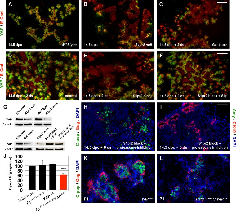
Fig 6. YAP mediates Gαi-dependent S1pr2 signalling and is essential for endocrine specification.
(A-B) Immunofluorescence analysis of embryonic pancreata at 14.5 dpc showed that YAP protein is specifically expressed in E-cadherin+ epithelial cells (A) and that it is dramatically reduced in S1pr2 nulls (B). (C-F) YAP expression is retained in the epithelium of 14.5 ALI embryonic pancreas explants cultured for 2 d (D) but dramatically reduced upon either S1pr2 block by 15 μM JTE013 (E) or Gαi inactivation by 10 μg/ml PTX (C). YAP expression levels are restored in JTE013-treated pancreata by 20 μM of S1p (F). (G) YAP protein levels detected by western blot were significantly reduced in S1pr2 null pancreata at 14.5 dpc and in 14.5 dpc ALI cultures subjected to S1pr2 blocking with 15 μM JTE013 for 2 d or subjected to Gαi inhibition by 10 μg/ml of PTX. YAP protein levels were restored in JTE013 treated pancreata by 20 μM of S1p or 1 uM of the proteasome inhibitor MG132. (H, I) Immunofluorescence analysis showed that C-pep+ and Gcg+ cells were restored in JTE013-treated 14.5 dpc + 6 d ALI cultures in the presence of 1 μM MG132 for the first 2 d (H). Amy+ cells, however, were not restored under these conditions (I) and CK19 expression remained high (I). Quantitations are provided in S7A Fig. (J–L) Inactivation of YAP in pancreas progenitors using the YAPfl/fl conditional allele, the TgPdx1CreERT2 driver, ip tamoxifen injections, and immunofluorescence analysis showed a nearly 40% reduction in the number of endocrine C-pep+ and Gcg+ cells compared to controls at P1. Scale bars, 80 μm (B-F, H, I) and 70 μm (K, L); ***p < 0.001, ns, not significant in reference to E14.5 samples (A) and wt embryonic pancreata; error bars show SEM with the exception of J, for which they show SD. For raw data, please refer to the S1 Data file.

