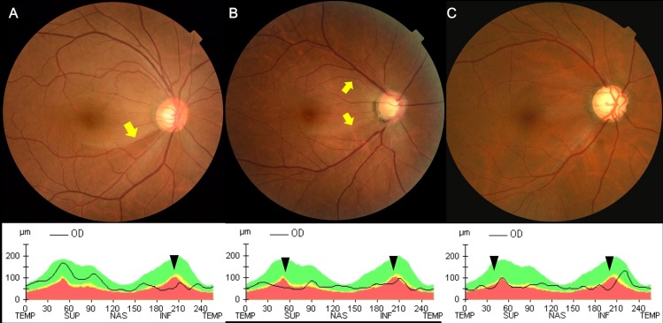
Fig 1. Fundus photographs and optical coherence tomography of representative patients in each group.
(A) In a patient with hemi-optic neuropathy, an inferotemporal defect in the retinal nerve fiber layer (RNFL) was evident on fundus photography and inferior thinning of the RNFL was apparent on the peripapillary RNFL thickness profile obtained using Cirrus HD optical coherence tomography OCT. (B) In a patient with bi-optic neuropathy, both superior and inferior RNFL defects were visible on fundus photography, and both RNFL thinnings were confirmed by OCT. The temporal margins of the RNFL defects evident on the fundus photographs of the above two patients, (A) and (B), are arrowed in yellow. (C) In another patient with bi-optic neuropathy, a diffuse RNFL defect was evident on fundus photography and a corresponding region of diffuse RNFL thinning was apparent on OCT. RNFL thinning based on OCT in the above three patients is indicated by black arrowheads.

