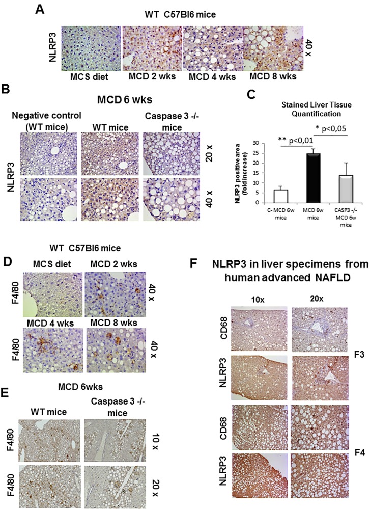
Fig 2. Immunohistochemistry analysis for NLRP3 and F4/80.
A,B. Immunohistochemistry analysis for NLRP3 on liver specimens from WT mice fed with MCS diet or MCD diet for 2wks, 4wks and 8wks (A) as well as WT mice and Casp 3-/- knockout mice fed for 6wks (B). C. Image analysis quantification for NLRP3 staining (Fig 2B) as evaluated with Imagej software in liver sections from WT or caspase 3 -/- mice fed a MCD diet for 6 weeks. D,E. Immunohistochemistry analysis for F4/80 on liver specimens from WT mice fed with MCS diet or MCD diet for 2wks, 4wks and 8wks (D) as well as WT mice and Casp 3-/- knockout mice fed for 6wks (E). F. Immunohistochemistry analysis for NLRP3 and CD68 on liver specimens from human NAFLD/NASH-related advanced fibrosis (F3 and F4). Original magnification as indicated.

