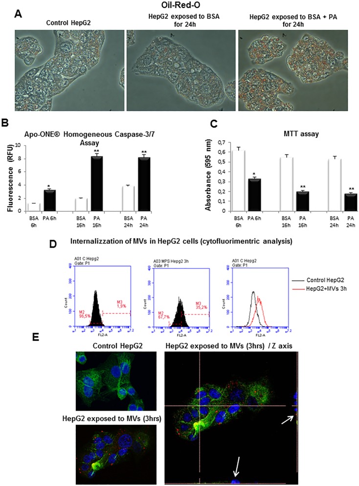
Fig 3. In vitro experimental model of lipotoxicity.
A. Red Oil-O staining in control HepG2 cells, HepG2 cells treated with BSA 1% or HepG2 cells exposed to palmitic acid 0.25mM in BSA 1% (BSA + PA) for 24hrs. B. Detection of Caspase-3/7 Activity in HepG2 cells treated with BSA 1% or PA 0.25mM for indicated times, analyzed by using Apo-ONE Caspase-3/7 Homogeneous Assay. C. Viability of HepG2 cells treated with BSA 1% or PA 0.25mM for indicated times, evaluated by MTT assay. D,E. Analysis of internalization of MVs in HepG2 cells by (D) flow cytometry or by (E) confocal microscopy: nuclei (blue fluorescence), MVs (red fluorescence) and cytoskeleton (F-actin, green fluorescence).

