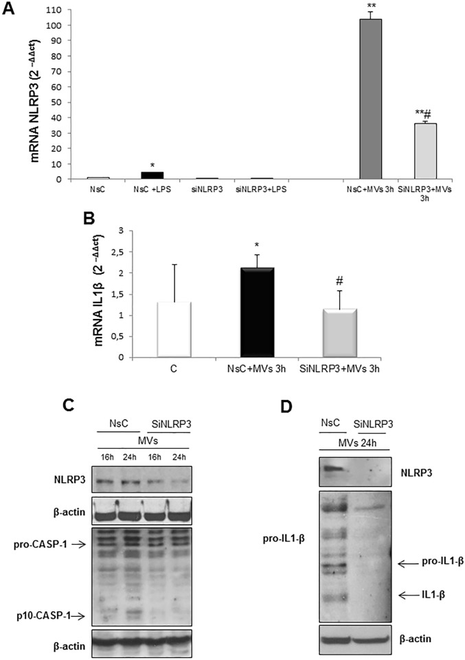
Fig 7. Silencing of NLRP3 in target cells prevents MVs-dependent inflammasome activation.
A. Quantitative real-time PCR (qPCR) of NLRP3 mRNA in HepG2 cells transfected for 72hrs with non-silencing siRNA (NsC) or specific siRNA for NLRP3 (siNLRP3) and exposed or not to MVs derived from HepG2 cells treated with PA 0.25mM for 24hrs. LPS 1mg/ml was used as positive control. B. Quantitative real-time PCR (qPCR) of IL-1β mRNA in HepG2 cells not transfected (C) and transfected for 72hrs with non-silencing siRNA (NsC) or specific siRNA for NLRP3 (siNLRP3) and exposed or not for 3hrs to MVs derived from HepG2 cells treated with PA 0.25mM for 24hrs. Data in graphs are expressed as means ± SEM (*p< 0.05 and ** p<0.01 vs related control HepG2 cells; # p<0.05 vs NsC+MVs) of three independent experiments. C,D. Western blot analysis of NLRP3 protein levels as well as of cleaved caspase-1 (p-10-CASP-1) and IL-1β in total extracts obtained by HepG2 cells transfected for 72hrs with non-silencing siRNA (NsC) or specific siRNA for NLRP3 (siNLRP3) and exposed for indicating time to MVs derived from HepG2 cells treated with PA 0.25mM for 24hrs. Equal loading was confirmed by re-probing membrane with β-actin.

