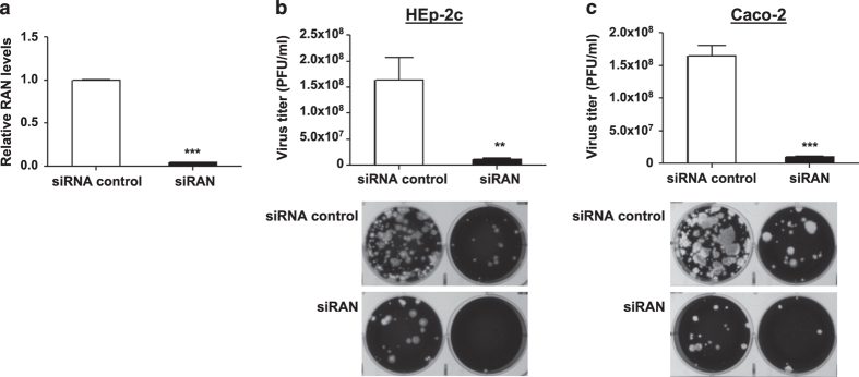
Figure 3. Silencing of RAN inhibits PV replication in HEp-2c and Caco-2 cells.
The cells were reverse-transfected with siRAN or siRNA control at a final concentration of 25 nM. After 48 h, (a) total RNA was isolated from HEp-2c cells and RAN mRNA level was determined by qRT-PCR. The data were normalized to the 18S rRNA gene and were expressed as fold change relative to siRNA control. The transfected Hep-2c (b) and Caco-2 (c) cells were infected with Sabin-2 strain at an MOI of 0.01 for 24 h, and then the virus titers were determined by plaque assay. The significant differences are compared with siRNA control. Error bars represent s.e.m. of quadruplicate. Each experiment was performed in quadruplicate and each experiment was repeated twice. The results were reproduced and the data in the figure were representative of 8 plates. **P<0.01, ***P<0.001.

