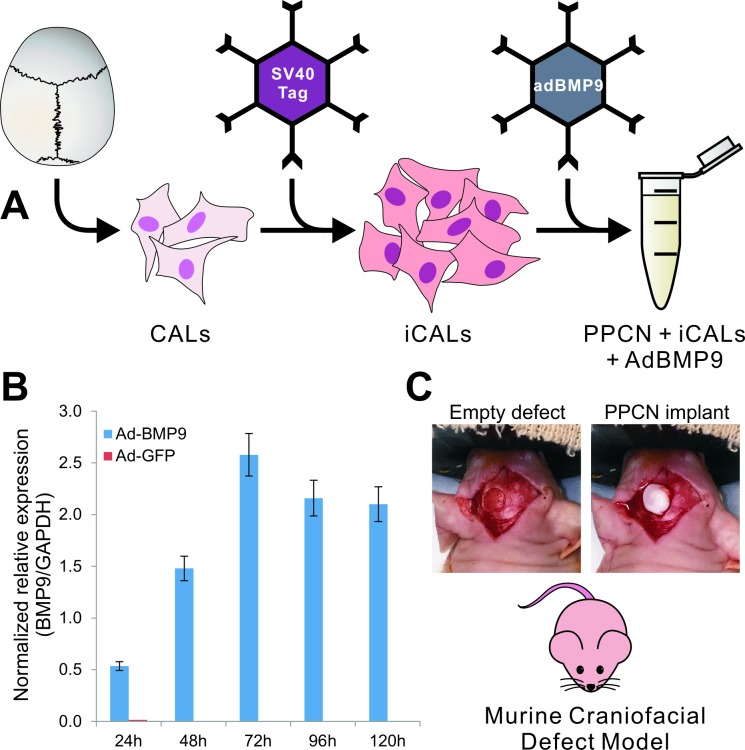
Fig 1. Schematic representation of the overall experimental design.
(A) Murine calvarial cells (CALs) were immortalized via retrovirally introduced SV40 large T antigen to produce iCALs. iCALs were then transduced with BMP9 via adenoviral vector and mixed with PPCN scaffolding material. (B) qPCR analysis demonstrating relative expression of BMP9 in iCALs infected with ad-BMP9 (blue bars) compared to control (ad-GFP)-infected cells (red bars). Gene transcript expression was normalized against GAPDH expression. (C) The mixture was subsequently tested in our murine craniofacial defect model. Four millimeter diameter full-thickness calvarial defects were created in the left parietal bone of 8-week-old male athymic (nu/nu) mice. The newly created empty defect reveals the underlying dura mater. PPCN alone or PPCN and adenovirally transduced immortalized calvarial cells were used to fill the defect site.

