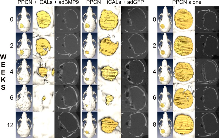
Fig 2. Time-course microCT imaging of the calvarial defects.
At 24–48 hours postoperatively, baseline microCT imaging was performed and analyzed to determine defect volume. Follow-up imaging and analysis was performed at 2, 4, 6, 8, and 12 weeks postoperatively to quantify residual defect volume and new bone ossification. Representative images are shown.

