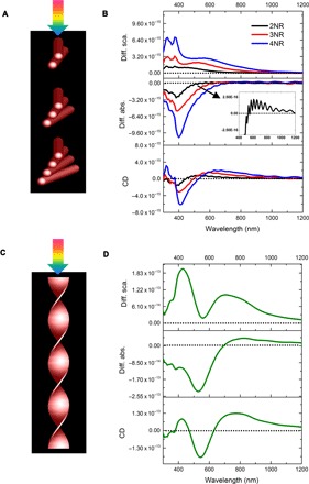
Fig. 7. Simulated nanorod deconstruction of a helix.

(A) Schematic of CPL irradiating a series of NR dimers (2NR), trimers (3NR), and tetramers (4NR). (B) Differential scattering (diff. sca.), differential absorption (diff. abs.) (inset shows a magnified view for dimer), and CD spectra for 2NR, 3NR, and 4NR. (C) Schematic of CPL irradiating onto a helix and (D) differential scattering, differential absorption, and CD spectra for the helix. Nanorod models are 312, 40, and 4 nm in length, diameter, and internanorod spacing, respectively, with a 10° dihedral angle.
