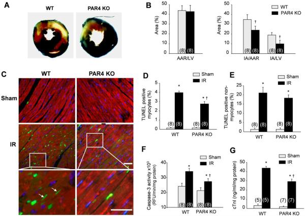
Fig. 2. PAR4 ablation reduces infarct size and cell apoptosis after acute IR.
Ischemia reperfusion (IR) was induced for 24 h. (A) Representative cross-sections were stained with triphenyl tetrazolium chloride and Evans blue to determine the extent of injury. (B) Quantification of infarct area (IA) vs. area at risk (AAR) after IR injury in the indicated groups. (C) LV tissue sections were assessed for apoptosis using TUNEL assay (green), tropomyosin (red), and DAPI (4',6-diamidino-2-phenylindole) (blue) staining. Arrows show TUNEL-positive myocytes. Scale bar, 40 μm. (D–E) The number of TUNEL-positive myocytes (D) and non-myocytes (E) in the reperfused area was expressed as a percentage of total nuclei detected by DAPI staining, n=8. (F) Quantification of caspase-3 activity in the LV using caspase-3 specific fluorogenic substrate, n=8. (G) Serum levels of cardiac troponin I (cTnI) after IR injury in WT (n=5) and PAR4 KO (n=7) mice. Values are presented as mean ±SEM, *P<0.05 vs. WT shams, †P<0.05 vs. WT IR.

